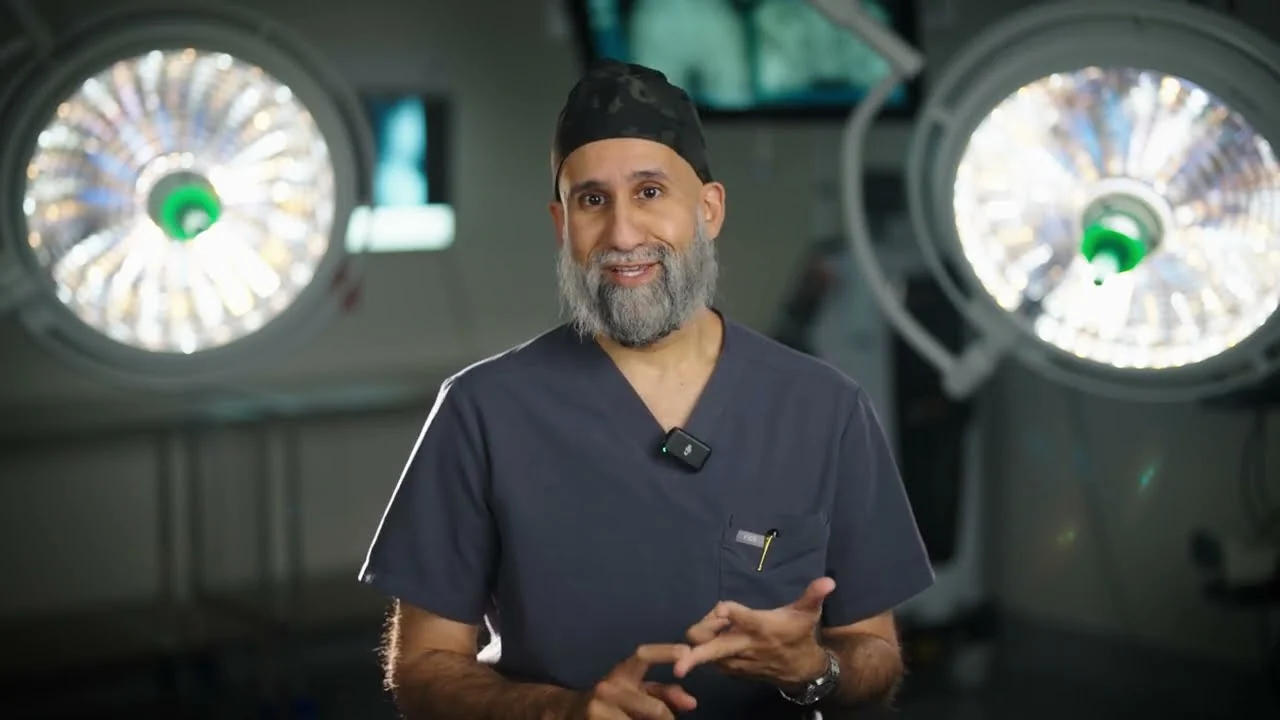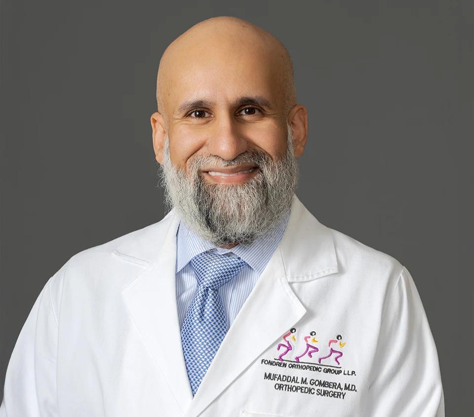
Medically reviewed by Mufaddal M. Gombera, MD, Board-Certified Orthopedic Surgeon.
Patellofemoral stabilization surgery stops a kneecap from dislocating by rebuilding the torn ligament that holds it centered and, when the anatomy demands it, correcting the track it rides in. Dr. Mufaddal Gombera is board-certified in orthopedic surgery with subspecialty certification in sports medicine and treats kneecap instability in athletes and active patients whose kneecaps have slipped more than once. If bracing and rehab have not stopped the dislocations, this is the operation designed to.
What Is Patellofemoral Stabilization?
Patellofemoral stabilization is the family of procedures that keep the kneecap riding in its groove instead of dislocating. The core operation is MPFL reconstruction, which rebuilds the ligament torn during dislocations. In knees whose bone shape keeps pulling the kneecap off track, a bone realignment procedure is added. The goal is straightforward: end the dislocation cycle described on our patellofemoral instability page before it costs the knee more cartilage.
MPFL Reconstruction, Explained Simply
The surgical procedure of medial patellofemoral ligament reconstruction involves the following steps:
Graft Selection and Harvest: Your surgeon will make a 4-6 cm skin incision over your knee, at the midpoint between the medial epicondyle and the medial aspect of the patella (knee cap). The underlying subcutaneous fat and fascia are cut apart to expose the adductor tendon. The tendon is then stripped using a tendon stripper and its free end is sutured. The diameter of the tendon graft is measured using a sizer.
Alternatively, a graft can be harvested from the quadriceps tendon.
Location of the femoral isometric point: The graft should be placed isometrically to prevent it from overstretching and causing failure during joint movements. A transverse hole measuring 2.5 mm is made through the patella. Then a small incision is made over the lateral side of the patella and a strand of Vicryl suture material is inserted through the hole. Over this strand, a 2.5 mm Kirschner wire (K-wire) is passed and then inserted into the bone besides the medial epicondyle.
An instrument called pneumatic isometer is inserted into the hole made in the patella and the Vicryl isometric measurement suture material is also passed along. The knee is taken through its full range of motion and any changes happening in the length between the medial epicondylar K-wire and the medial aspect of the patella is recorded on the isometer. The position of the K-wire will be adjusted until no deviations are read on the isometer during full range of motion. Once the isometric point is identified, a tunnel is drilled starting from the insertion of the adductor tendon until the isometric point is reached. The graft is pulled through this tunnel, then exits at the medial condyle and again passed through another tunnel that is made through the patella.
Correct tension: The tension is set in the graft with your knee flexed up to 90ºand the tension should be appropriate enough to control lateral excursion.
Secure fixation: After bringing the tendon graft from the medial to the lateral side through the bone tunnel, it turned onto the front surface of the patella where it is sutured.
Avoid condylar rubbing and impingement: After graft fixation, the range of motion is checked to make sure there are no restrictions in patellar or knee movements. The graft should not impinge or rub against the medial femoral condyle. If it is detected, the graft is replaced into proper position.
Post-operative care
The MPFL, the medial patellofemoral ligament, is the seatbelt on the inner side of the knee that stops the kneecap from sliding outward. Dislocations tear it, and once torn it usually heals stretched and unreliable. Reconstruction replaces it with a tendon graft anchored between the inner edge of the kneecap and a precise point on the thighbone.
Precision is the entire game in this operation. The graft must be attached at exactly the right spot and set to exactly the right tension: tight enough to check the kneecap, loose enough that it does not press the kneecap into the groove and create new pain. Before the reconstruction, Dr. Gombera examines the joint arthroscopically and treats what the dislocations left behind, including loose fragments and cartilage damage on the back of the kneecap.
When Bone Procedures Are Added
Soft tissue alone cannot out-pull bad bony alignment, so some knees need the track corrected, not just the seatbelt replaced. The workhorse bony procedure is the tibial tubercle osteotomy, or TTO: the small bump on the shinbone where the patellar tendon attaches is shifted a short, measured distance, usually inward, and fixed with screws so the tendon pulls the kneecap straighter. It is added when imaging shows the attachment sits too far outward or the kneecap rides too high. For the rare, severely shallow groove, deepening procedures exist; whether that step is appropriate is a candid, case-by-case conversation.
Discover Your Ortho Options
What Surgery Involves
Stabilization is outpatient surgery under anesthesia. Dr. Gombera harvests or prepares the tendon graft, inspects the joint through the same technique used in knee arthroscopy, addresses cartilage damage, then anchors the graft to rebuild the MPFL, adding the bony realignment when the plan calls for it. You go home the same day with a brace and crutches, and physical therapy begins within days, because early guided motion prevents the stiffness that used to plague kneecap surgery.
Risks, Stated Honestly
The important ones: recurrent instability (uncommon, but no operation reduces the risk to zero), stiffness, pain from a graft tensioned too tightly, hardware irritation after bone realignment, and the standard surgical risks of infection and blood clots, which are rare. Choosing the right combination of procedures for your anatomy, rather than one default operation for everyone, is the main protection against most of these, and it is how Dr. Gombera plans every case.
Recovery Timeline
- Day of surgery: Outpatient surgery. Home the same day with a brace and crutches.
- Weeks 1 to 6: Brace for walking while the graft protects itself. Early motion and quadriceps activation in physical therapy start right away.
- Weeks 6 to 12: Wean out of the brace. Progressive strengthening and balance work.
- Months 3 to 4: Running as strength and control return.
- Months 4 to 6: Sport-specific training and return to sport for most athletes.
Frequently Asked Question
What does MPFL reconstruction actually do?
It rebuilds the medial patellofemoral ligament, the seatbelt on the inner side of the knee, using a tendon graft anchored between the kneecap and thighbone. The new ligament checks the kneecap’s tendency to slide toward the outside of the knee.
Will surgery guarantee my kneecap never dislocates again?
No honest surgeon guarantees that. What the operation reliably does is drop the risk of another dislocation dramatically, especially when bony risk factors are corrected at the same time. The large majority of patients never dislocate again.
When is a bone procedure added to MPFL reconstruction?
When the anatomy that caused the dislocations would doom a soft tissue repair working alone: a tendon attachment that pulls the kneecap far outward or a kneecap riding too high. In those knees, a tibial tubercle osteotomy realigns the pull.
How long will I wear a brace after surgery?
A brace is used for walking during roughly the first six weeks while the graft is protected. Physical therapy starts almost immediately, because early motion prevents stiffness without endangering the reconstruction.
When can I get back to sports?
Most athletes return to sport around four to six months after surgery, once strength and control match the other leg. Knees that also had bone realignment may run a few weeks behind that pace.
Is the surgery different for teenagers?
It can be. In younger patients with open growth plates, graft placement and any bone work are planned around the growing bone.
Schedule a Kneecap Stabilization Consultation in Houston
Dr. Gombera performs patellofemoral stabilization at Texas Orthopedic Hospital (7401 Main St), with clinics at Memorial (8731 Katy Freeway) and Baytown (7718 Garth Rd). Same-week consultations are available.